

|
Presentation Biology Living habits Infestation Control methods |
||
|
|
||
|
|
Author: François DROUET. |
|
|
|
||
|
The egg (maturation, incubation)
The description of the egg of Silba adipata McAlpine, accompanied by observations of unhatched eggs and empty eggs envelopes (chorions), is the subject of a specific chapter. In this chapter, I give information and I provide observations about the maturation and the incubation of the egg. According to the following plan: female reproductive system, egg maturation, the egg just after laying, revealing the yolk under a stereomicroscope, incubation.
FEMALE REPRODUCTIVE SYSTEM
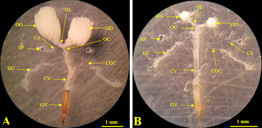
MODEL The female reproductive organs of insects have a common structure. Until 2025, no photographs of the reproductive system of Silba adipata McAlpine had been published. The closest example found in the specialized literature, and published in this chapter, was that of Bactrocera dorsalis Hendel, a polyphagous fruit fly belonging to the Tephritidae family. Reference: GOMINA M., 2015, Contribution à la connaissance des mouches des fruits (Diptera : Tephritidae) et de leurs parasitoïdes dans le sud du Togo, doctoral thesis in life sciences, developmental biology, applied entomology option, University of Lomé (Togo). In 2025, we included in this chapter the first published photograph of the female reproductive system of Silba adipata McAlpine (see below). However, we felt it was useful not to remove the work of M. GOMINA, which is of great educational value. The reproductive system of sexually mature females (photograph A, below) consists of two ovaries, left (OG) and right (OD), each composed of numerous ovarioles (linear series of developing oocytes). The ovaries continue into two short lateral oviducts (OL), merging into a common oviduct (OC), or vagina. The vagina is connected to the ovipositor (OV) by a vaginal duct (CV). Two spermathecae (SP) open into the vagina via ducts (CS). They receive and store sperm during mating, and release it when the egg passes into the vagina. Two elongated colleteric glands (GC) also open into the vagina via their ducts (CGC). They produce an adhesive substance that fixes the egg to the support where it is laid. The opening located at the level of the ovipositor has a triple role: anus, insertion point of the aedeagus (male copulatory organ), exit point of the eggs. The reproductive system of females dissected just after emergence (photo B, below) has the same organization as that of sexually mature females. But it is characterized by very small ovaries, which are made up of ovarioles that are difficult to observe, in which no developing oocytes are visible.
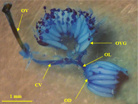
Bactrocera dorsalis Hendel: female reproductive organs, after (A) and before (B) sexual maturation. The photograph below shows the detail of the ovaries of a sexually mature female. These have been stained with methylene blue to make them more visible.
Bactrocera dorsalis Hendel: ovaries of a sexually mature female (left ovary unfolded to observe the ovarioles).
CASE OF SILBA ADIPATA McALPINE In 2025, DIAZ-DEL-CASTILLO R. et al. published in the Insects journal an article relative to the sexual development of Silba adipata McAlpine. Reference: DIAZ-DEL-CASTILLO R.,
CÓRDOVA-GARCIA G.,
PÉREZ-STAPLES D., BIRKE A.,
WILLIAMS T., LASA R., 2025, Sexual
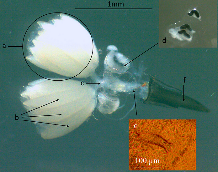
Development of Silba adipata (Diptera: Lonchaeidae): Effects of Diet, Ultraviolet Light and Fig Latex, Insects 16, no. 5: 495. This article includes a photograph showing the female reproductive system, that we provide below (authorized reproduction, Open Access license).
Silba adipata McAlpine: female reproductive organs. The photograph is accompanied by a description that we do not reproduce in extenso, limiting ourselves to mentioning the differences with the previously seen model. The authors cite (without noting them on the photograph) the lateral oviducts connecting the ovaries to the common oviduct. Concerning the latter, they consider that it continues to the ovipositor (not calling the vaginal duct the wider part of this canal). Concerning the spermathecae, they count three (a pair, and one placed individually), and specify that these are black, sigmoidal, and covered with a translucent whitish capsule (at the top, on the right, in the photograph, there is an enlargement of the spermathecae freed from their capsule). The colleteric glands and their ducts are not cited, nor highlighted in the photograph. And the authors highlight the presence, quite low in the common oviduct, of a sclerotized structure, with a series of comb-like projections along its edges, and appearing to have an opening oriented towards the ovipositor. They specify that its location and morphology suggest that it is likely a signum (e), of which they provide an enlargement in orange. The authors indicate that the signum is a sclerotized structure present in the female reproductive tract of certain arthropods, which plays a significant role in reproduction, although its shape and size can considerably vary among species. But they do not specify what could be the exact role of the signum in the specific case of Silba adipata McAlpine.
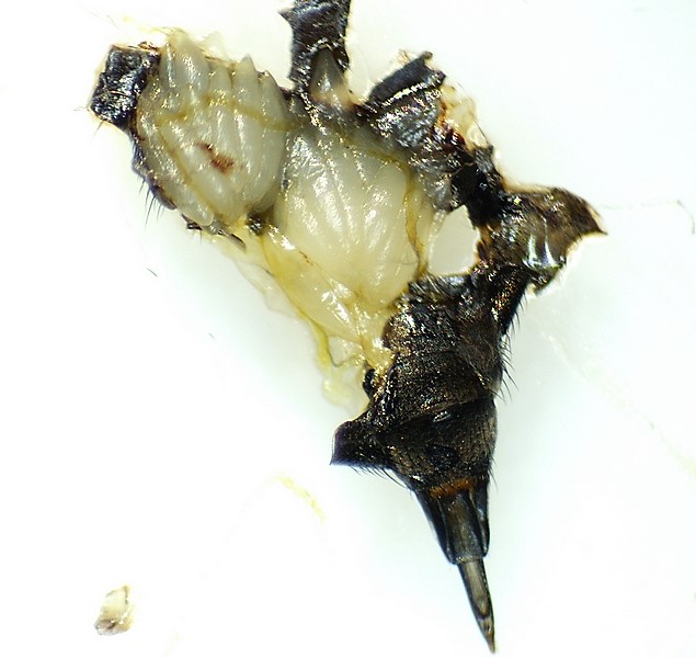
OURS OBSERVATIONS We do not intend to conduct a detailed study of the female reproductive system of Silba adipata McAlpine. But we have opened the abdomens of more than 200 females of the species, particularly to study the population dynamics of mature females during the period of intense attacks on the immature figs, and we observed three stages of female sexual development: immature female, female with eggs maturation more or less advanced but incomplete, mature female. When the eggs maturation is advanced but incomplete, the eggs are clearly visible but insufficiently chorioned. Consequently they cannot be effectively counted because they burst easily during handling, turning into a whitish mush. Below is a photograph of the ovaries of a Silba adipata mature female.
Silba adipata McAlpine : ovaries of a mature female.
MATURATION (FORMATION) OF THE EGG
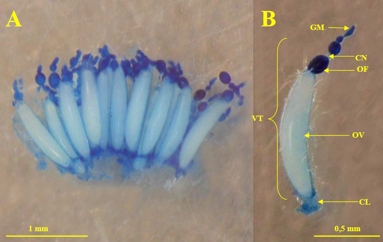
Maturation of the egg is the phase during which it forms within the female reproductive system, more precisely in an ovariole. Each of the ovarioles of a sexually mature female (photographs below) is an epithelial tube in which we observe a growth zone: the germarium (GM), a maturation zone: the vitellarium (VT), and a calyx (CL), or pedicel (which opens into the lateral oviduct). The vitellarium is made up of a linear series of forming oocytes, which ends with a mature chorionated oocyte (OV), that is to say provided with its external envelope which will protect it after egg-laying (chorion). Each forming oocyte (OF) is accompanied by nurse cells (CN), called trophocytes, which persist until the complete formation of the chorion.
Bactrocera dorsalis Hendel: ovarioles of a sexually mature female, stained with methylene blue to make them more visible. Détails about egg maturation (text and figures below) are provided by the insect development biology course of Sihem AGUIB. Reference: AGUIB Sihem, 2020, cours
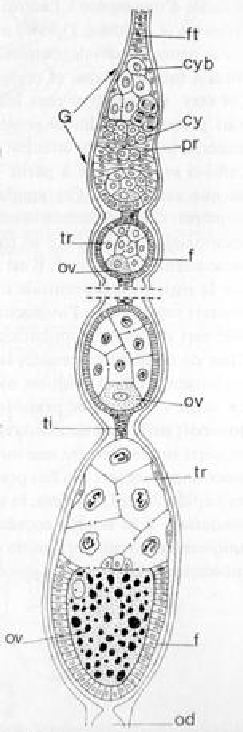
Biologie du développement des insectes (L3 entomologie), Frères Mentouri University, Constantine (Algeria). There are three types of ovarioles in insects. Diptera (including Silba adipata McAlpine) are among the insects that have the most complex of the three types of ovarioles, known as meroistic and polytrophic (figure below).
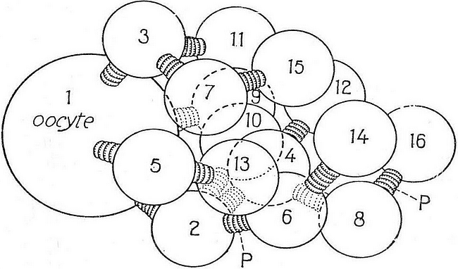
Meroistic and polytrophic ovariole in Drosophila (M. R. CUMMINGS and R. C. KING). The epithelial tube constituting the ovariole runs from the terminal filament (ft) to the pedicel (od), the latter leading to the lateral oviduct. Oogonial differentiation takes place in the germarium (G). The cystoblast (cyb) from a primary oogonia undergoes a specific number of mitoses and gives rise to a cyst of cells, called cystocysts (cy), for example 16 cells in Drosophila. All of these cells will be enclosed in a follicle. One of these cells (the most posterior inside the follicle) becomes an oocyte (ov), which enters meiotic prophase, then the period of vitellogenesis. The other cells evolve into trophocytes (tr). The germarium also contains, at its base, prefollicular cells (pr). Upon entering the vitellarium, each oocyte, along with the associated trophocytes, is enveloped by a follicle consisting of follicular cells (f). The follicles are separated by clusters of interfollicular tissue (ti), which are modified follicular cells. The trophocytes accompanying the oocytes as they descend into the vitellarium first increase in size and become polyploid, then they disintegrate, their cytoplasm flowing into the oocyte.
Spatial representation of the connections between the 16 cells of a follicle in Drosophila.
THE EGG JUST AFTER LAYING
Silba adipata McAlpine: egg under an ostiolar scale of an immature fig - 'Bellone' variety.
Silba adipata McAlpine: egg under an ostiolar scale of an immature fig - 'Bellone' variety. Details on the contents of the egg just after laying (text and figure below) are provided by the insect development biology course of Sihem AGUIB. Reference: AGUIB Sihem, 2020, cours Biologie du développement des insectes (L3 entomologie), Frères Mentouri University, Constantine (Algeria). The newly laid egg is filled with yolk, a nourishing substance used for the development of the larva. The yolk is surrounded by a thin translucent membrane, the vitelline membrane (mv). And the egg is surrounded by a thick protective envelope, the chorion (ch), which has been secreted by the follicular cells in the ovariole. The yolk is made up of yolk globules (gv) of diverse chemical nature (proteins, etc.), located in a cytoplasmic network, the cytoplasmic reticulum (ret). This is connected to a thin layer of peripheral cytoplasm devoid of yolk globules, the periplasm (pe), or cortex. A special plasm, or oosome (oos), rich in ribonucleoproteins, is formed at the posterior pole of the egg. It is involved in the larva formation process during the egg incubation phase. In addition to the yolk, the egg contains a fertilization nucleus, located in a thickened area of the yolk mass, called the maturation plasm (pm). When the oocyte left the ovariole to pass into the genital tract, meiosis was blocked in metaphase I. When the oocyte passed in front of the opening of the spermatheca, spermatozoa were released and entered the oocyte through the apical openings of the latter, the micropyles (m). Meiosis completes rapidly in the maturation plasm. Fusion of the two nuclei (amphimixis) restores the diploid number of chromosomes.
Sagittal section of the egg just after laying (example of the Drosophila egg, according to A. HAGET).
REVEALING THE YOLK UNDER A STEREOMICROSCOPE
When I counted eggs in immature figs under a stereomicroscope, I noticed that the chorion came apart quite easily when I scraped the unhatched egg with one of the tips of the dissecting forceps, while holding it with a rounded-tipped pick. It then appears, either the yolk enclosed in the vitelline membrane, which turns out to be surprisingly solid (egg recently laid, or not fertilized), or a larva (egg laid for a longer time, incubation well advanced). In some cases, I use the contents of the egg to determine the uniqueness or the plurality of clutches in a fig.
Silba adipata McAlpine: 4 eggs, 1 of which with the yolk fully revealed after scraping the chorion; on its left, the removed chorion.
Silba adipata McAlpine: egg with the yolk partially revealed, after scraping the chorion.
INCUBATION
Incubation of the egg is the phase during which the larva forms inside it. It begins as soon as the egg has been laid, by division of the fertilization nucleus located in the maturation plasm. According to the observations of F. SILVESTRI, the incubation of a Black Fig Fly's egg lasts 8 days in April, but only 3 days in summer. It therefore takes 3 to 8 days, depending on the season, for the larva to hatch from the egg. Reference: SILVESTRI F., 1917, Sulla Lonchaea aristella Beck. (Diptera : Lonchaeidae) dannosa alle infiorescenze e fruttescenze del caprifico
e del
fico, Bollettino del Laboratorio di Zoologia Agraria in Portici, vol.12, pp. 123 -146.
Silba adipata McAlpine: 1 larva and 3 chorions under an ostiolar scale.
|
||
